Injuries and fractures of the upper extremities
3 The value of the use of physiotherapy for fractures
5 Mechanisms of action of the applicator
6 Application therapy for injuries and fractures of the upper limbs
The structure of the bones
The human skeleton consists of 206 bones, different in shape, structure and size. It is represented by a wide variety - from wide and flat bones of the cranial vault to long tubular bones of the limbs.
The functions of the bones are to maintain the shape of the body, provide support for it and participate, together with the muscles, in movement. The skeleton protects vital organs from damage. The skull protects the brain, organs of vision and hearing, and the chest protects the heart and lungs.
Bone tissue is a specialized type of connective tissue with a high mineralization of intercellular organic matter containing about 70% inorganic compounds, mainly calcium phosphates. More than 30 microelements (copper, strontium, zinc, barium, magnesium, etc.) have been found in bone tissue, which play an important role in metabolic processes in the body.
Bones are the main storage of the mineral calcium, they contain 99% of its reserves. Calcium is not only a building material for bones and teeth, but also takes part in many metabolic processes in the body, such as nerve conduction, blood clotting, muscle contraction.
The structure of the bone (Fig. 1). Each bone is covered on top with a fibrous connective tissue film periosteum, which is permeated with blood vessels that deliver nutrients and hormones to bone cells. As well as nerves that transmit information about pain and the position of the bone in space. Under the periosteum is a compact bone substance - densely packed hard tissue, and deeper - a cellular spongy substance.
The composition of the bone. Bone tissue is formed by a dense framework of collagen protein fibers (an organic substance that provides elasticity), which is filled with crystals of calcium salts (a mineral substance that gives strength to bones). Collagen is a strong and flexible material that is the basis of most connective tissues, such as bone, cartilage, fibrous (in the skin, tendons, ligaments).
Despite the high degree of mineralization, in the bone tissues there is a constant renewal of their constituent substances, constant destruction and creation, and adaptation to changing operating conditions. The morphological and functional properties of bone tissue change depending on age, physical activity, nutritional conditions, as well as under the influence of the activity of the endocrine glands, innervation and other factors.
Bone cells. Bone tissue consists mainly of collagen and calcium salts, but it is a living structure that is constantly changing, breaking down and renewing with the participation of special cells: osteoblasts, osteocytes, osteoclasts.
Osteoblasts produce a protein that forms the collagen frame of the bone and calcium ions, i.e. synthesize the intercellular substance of the bone. They are located under the periosteum.
Osteocytes. When osteoblasts are immured in the formed bone material, they reduce their activity and turn into osteocytes with long processes. These are very numerous cells of the bone tissue, which provide it with a metabolism.
Osteoclasts. They work to remove damaged tissue in the bone to form new tissue, or to release calcium from the bone that is needed elsewhere in the body. For these purposes, the cells destroy the intercellular substance of the bone or excrete calcium with the help of acid and enzymes.
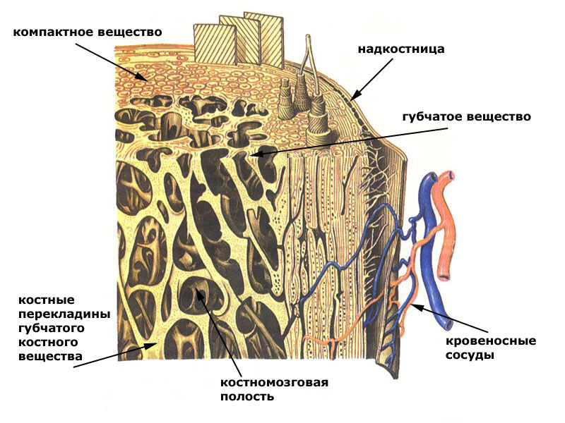
Fig.1
Fracture healing
After a fracture, the body immediately tries to restore the bone tissue to its original state. It takes six weeks to several months for the bone to heal completely.
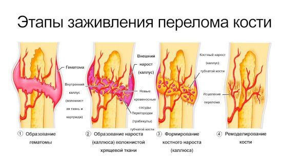
Immediately after the fracture, bleeding begins from the damaged vessels of the periosteum. A blood clot called a hematoma forms. The blood coagulates, stops flowing out, and fibrous connective tissue gradually forms in place of its clot. After a few days, tough tissue called calluses develops at the sites of the hematoma. Cells called fibroblasts begin to produce fibers of collagen, the main protein in bone and connective tissue. Chondroblasts then begin to produce fibrous cartilage. This tissue stiffens calluses by closing the gap between parts of the bones. This process takes about three weeks.
Bone callus after a fracture.
The osteoblasts then begin to produce bone cells, turning calluses into calluses. This bone shell provides the necessary protection for 3-4 months before the bone goes through the final stage of healing. A sufficiently large amount of time must elapse before the bone is ready to take on the full load. Osteoclasts and osteoblasts will work for several months to turn the callus into a full-fledged bone tissue. These cells also reduce the bulge, returning the bones to their original appearance. Circulation improves along with an influx of nutrients such as calcium and phosphorus, which strengthen bones. Even the most minor fractures require medical attention so that the healing process goes as quickly as possible.
The value of the use of physiotherapy for fractures
After a fracture, it is necessary first of all to restore the integrity of the damaged bone. For this purpose, methods of traction, immobilization and surgical intervention are used.
But no less important is the return of mobility, innervation, trophism of the injured limb. Medicine has many methods aimed at rehabilitation after fractures, and one of the most significant is physiotherapy.
It is difficult to overestimate the importance of physiotherapeutic procedures at the stages of treatment and rehabilitation after fractures. As part of complex therapy, they contribute to recovery and return to working capacity, alleviate the patient's condition. The main tasks of physiotherapy treatment:
- prevention of muscle atrophy and the formation of contractures;
- reduction of puffiness;
- elimination of pain syndrome;
- improvement of microcirculation and tissue nutrition;
- prevention of infectious and inflammatory processes in open fractures.
A properly selected set of procedures not only promotes recovery, but also reduces the drug load on the body.
The choice of specific methods of physiotherapy treatment directly depends on the stage of bone tissue regeneration.
The following types of physiotherapy are shown: UHF, Magnetotherapy, electrophoresis, massage, Lyapko application therapy, etc.
Application therapy Lyapko
Lyapko's applicators in various modifications (plates, rollers, applique belts, applique tapes) are an original, powerful device with many health-improving therapeutic possibilities. Their action is based on the principles of traditional Chinese medicine - superficial multi-needle acupuncture, as well as on the general physiological mechanisms of life.

Mechanisms of action of the applicator
The high healing effect of Lyapko applicators is due to a combination of intense reactions:
- reflex-mechanical;
- galvano-electric;
- immunological.
The clinical effects of the method of multi-needle therapy are manifested in analgesic, antispasmodic, anti-inflammatory, anti-edematous, neurotrophic and immunomodulatory effects. Also in the regulation of the functions of the autonomic nervous system, the normalization of the processes of excitation and inhibition in the central nervous system.
Application therapy for injuries and fractures of the upper limbs
If there are metal plates, pins, fragments in the patient's body, then any type of electrophysiotherapy is contraindicated for them and the only means of physiotherapeutic treatment is the Lyapko applicator. Lyapko's application therapy can be used from the first days of an injury or fracture, it is combined with all types of medical and physiotherapeutic treatment.
How to work with the applicator, application zones
In 90% of cases, it is necessary to act on the pain zone, and to increase efficiency on additional and auxiliary zones. It is always necessary to include the main zone (the region of the spine) in the general recipe.
Additional and auxiliary zones should be used when, for a number of reasons, it is impossible to influence the main zone (gypsum is applied, the wound surface). To enhance the therapeutic effect, it is advisable to include in the formulation the effect on the symmetrical zones of the healthy side.
The main zones are located on the back surface of the trunk, head, neck. The main ones are named due to the fact that the skin areas on both sides of the spine and directly above the spine are closest to the exits of the roots of the cranial and spinal nerves and other structures.
Auxiliary zones: front surface of the trunk, head and neck.
Additional zones: zones of the skin of the lower and upper extremities, which are secondary (peripheral) in relation to the (central) structures of the spinal cord and brain.
General recommendations
When applying plaster in the first days, there is often a strong swelling, increased pain at the fracture site, leading to the fact that you have to cut the plaster. First of all, you need to act with any flat applicators on the cervical-collar zone of the spine, from where the innervation to the limb comes from - the main zone, if there is access there. It is also recommended to act on all free surfaces of the hands above and below the fracture site (plaster cast), as well as on a healthy arm, focusing on the symmetrical fracture site, this reflexively reduces swelling and pain on the injured limb. The impact can be carried out both with a flat applicator, and with a “Large Roller M” , “Universal Roller M” . You can also use static-dynamic applicators:"Magic tape "Health" , belt "Universal M" , belt "Baby" . They need to be fixed on the limb for a longer time from 30 to 60 minutes 2-3 times a day.
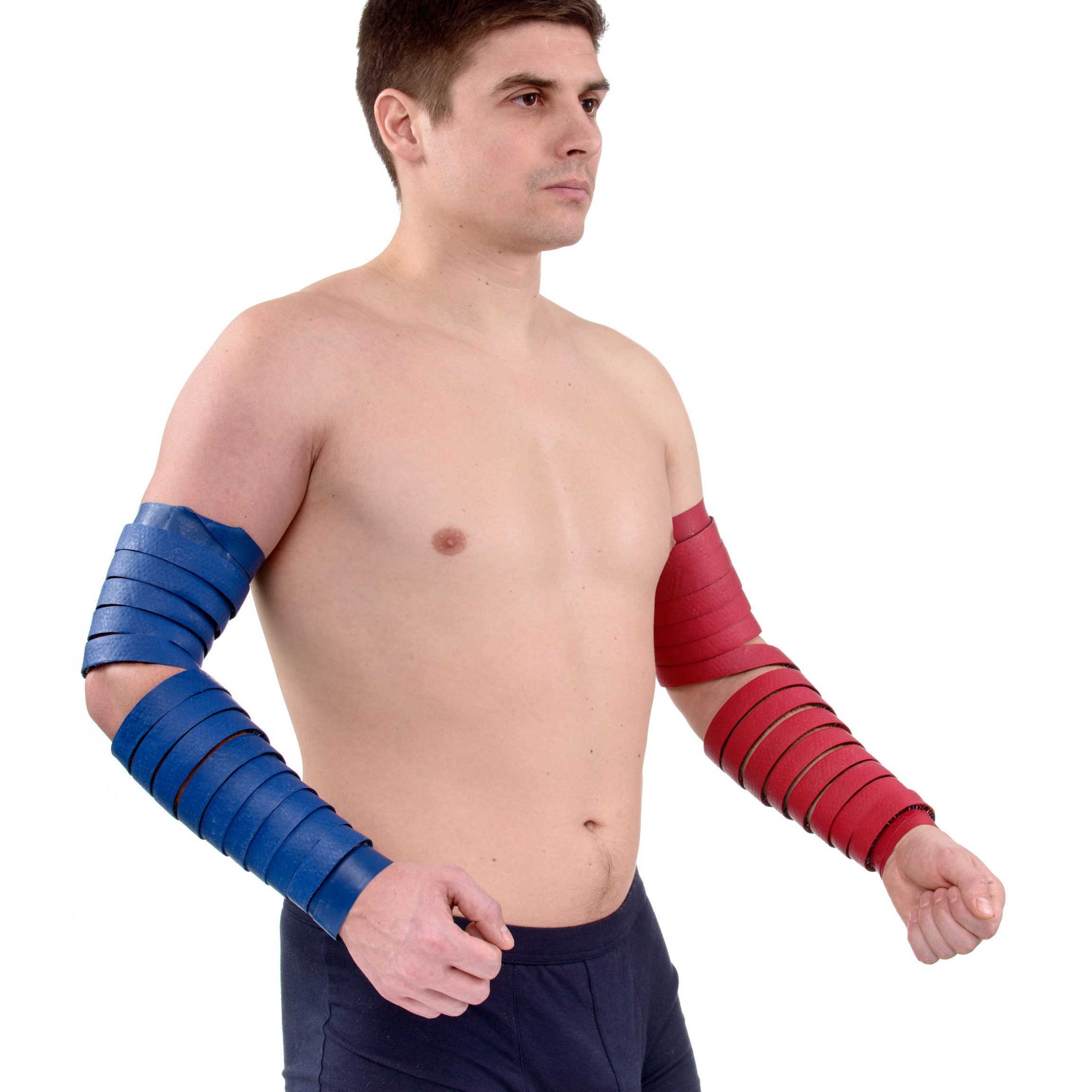
For injuries - the same technique. If the skin over the injured area of the limb is not damaged, then you can act on the site of injury. In case of bruises, to avoid the occurrence of hematomas, you can fix any flat applicator, application tape or roll it with a roller to this place.
Application zones (Fig. 2): main 2, 3; additional 4, 13; auxiliary 20, 22, 24, (28, 30).
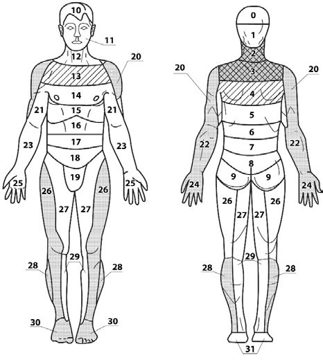
Fig.2
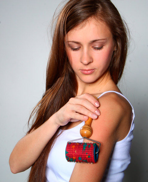
Also roll with a roller, " Needle ball " fingers on a healthy hand and on an injured one. The duration of exposure on the side of the fracture is 2 times longer than on the healthy arm.
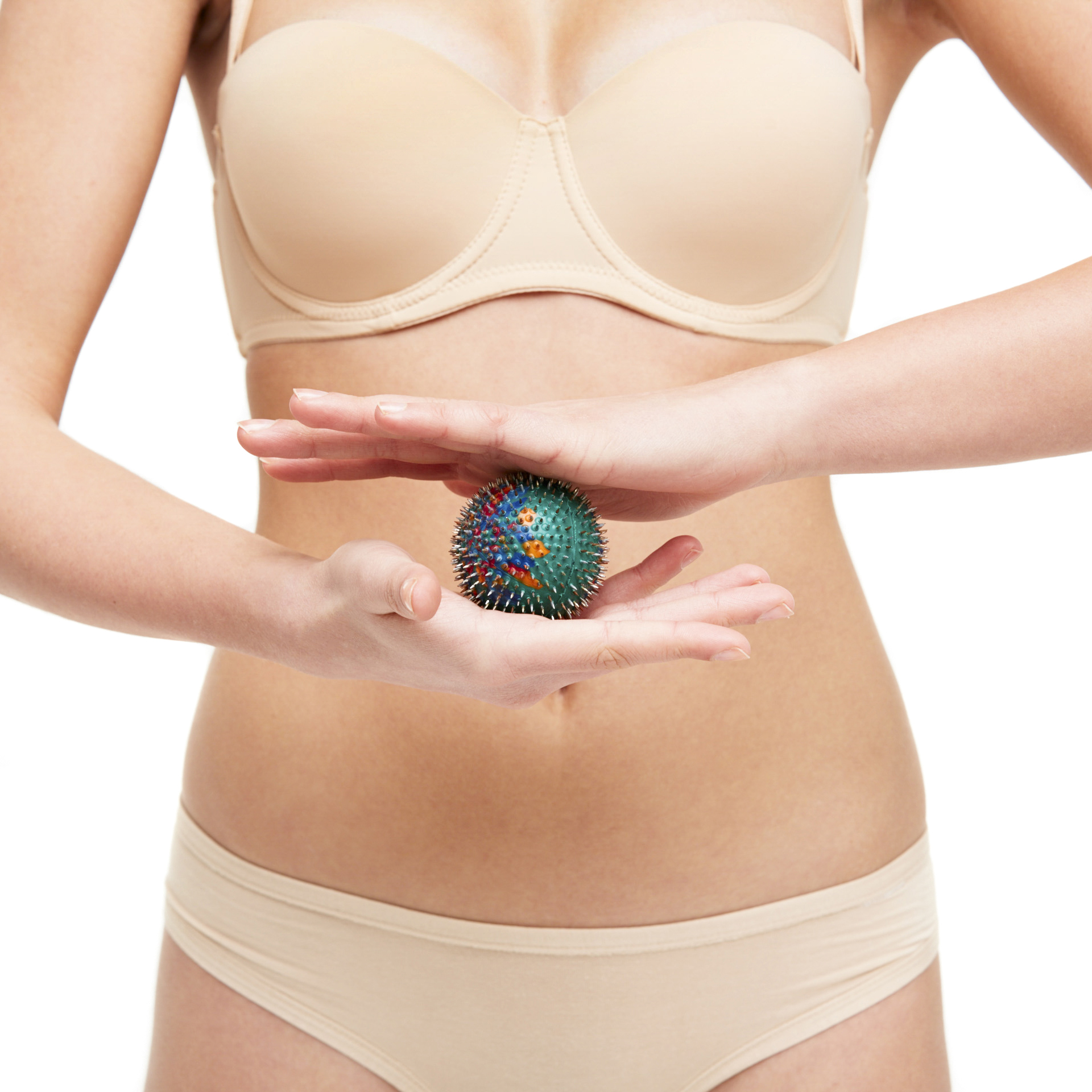
For example, exposure with static applicators for 15-20 minutes on the neck-collar area and on a sore hand, 5-7 minutes on a healthy hand.
Additional recommendations. A good result gives a short-term effect on the symmetrical zones of a healthy limb, the zones of correspondence on the hands and feet, used in Su Jok therapy (Fig. 3)
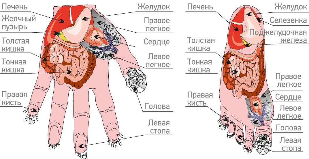
Fig.3
If pain zones and zones of unusual sensitivity are detected using the “Facial Roller M” or “Universal Roller M” , small “Kraplink” applicators should be fixed on them for 1–2 hours.
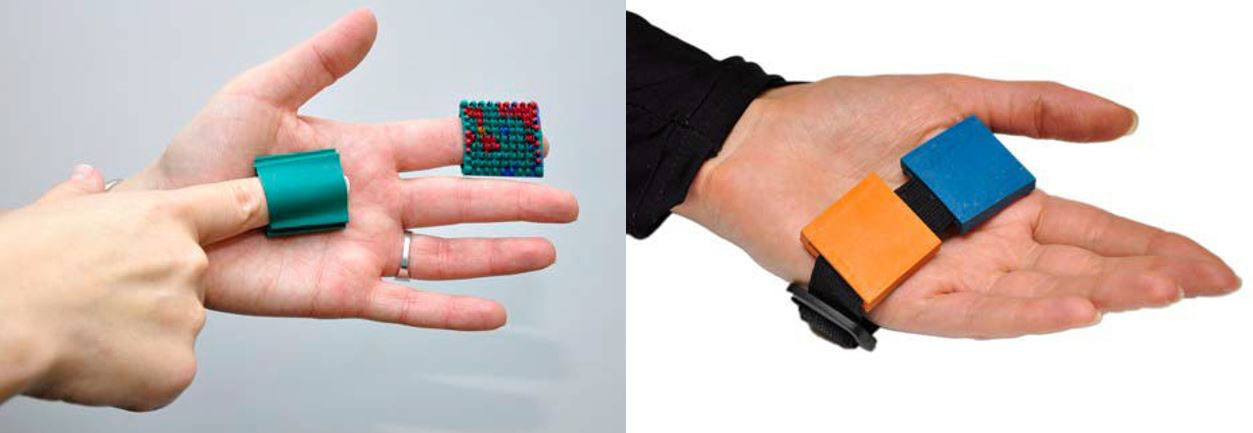
The composition of the applicator needles includes copper and zinc, which stimulate and accelerate the healing and callus formation processes.
In the old days, healers, in the treatment of fractures, practiced this method: with a small file from an old copper coin, copper powder was planed. A little of this powder (1/10 gram), stirred in milk or sour cream, or rubbed with egg yolk and given to the patient inside, 2 times a week. This accelerated the healing of fractures.
You need to eat food rich in vitamins and microelements such as: manganese, copper, zinc, phosphorus, calcium, magnesium, vitamins B6, B9, B12, C, D, K. All these elements help to absorb calcium and protein - from which the body and will build bone tissue.
It is advisable to use the mummy inside and outside (in solution) on the fracture area.
In the early days, with pathology of muscles, bones and joints, rest should be observed, but in the future, physical activity should be gradually increased. If the muscles do not work, they quickly weaken and atrophy. Without load, the work of the ligamentous-articular apparatus is disrupted, which lengthens the process of recovery and recovery after an injury, and increases the likelihood of future injuries.
After the cast is removed, the muscles are always painfully tense, require gradual stretching, and the joints need to be “developed”. After removal of the plaster, pain can appear in almost all parts of the back, since immobilization of both the upper and lower limbs after fractures leads to a gross violation of the stereotype of movements of the whole body and the appearance of pronounced asymmetries of the body. Therefore, to prevent the appearance of muscle pain in the back, from the first days of the fracture, flat applicators should be applied to the back for 20-30 minutes in the evening, roll the back with the "Big Roller M" , "Universal Roller M" in the morning and afternoon for 3-5 minutes (until a uniform pink color appears). Do a back massage using the Pharaoh massager .

After removing the plaster, we continue to work on the diseased hand and on the healthy one. In case of fractures in the wrist joint, it is recommended to wrap the hand with the Magic Ribbon Health applicator , twist it in the palm of your hand and squeeze it with a roller or Needle Ball applicator . Holding the ball in the hand, rotate it (turns).

